
RETINAL DETACHMENT
General Description
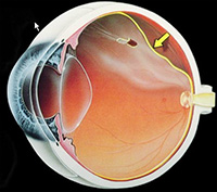 The detachment of the retina is an ophthalmologic emergency. The retina is a thin layer of light-sensitive tissue on the back wall of the eye which sends the images as electrical impulses to the brain. If the retina is detached from the back wall of the eye, it loses its blood flow and the tissue begins to die.
If treated in time, vision can be recovered. If surgery is delayed in the case of retinal detachment, the vision can be completely lost.
Causes
Contrary to popular belief, retinal detachment is not always associated with trauma.
Often there are pathological conditions which predispose the patient. These conditions cause damage to certain areas of the eye, most commonly in the periphery of the retina.
These weak zones are called retinal degenerations. It is important to undergo a complete ophthalmologic evaluation in order to diagnose and treat these degenerations appropriately.
Retinal detachments are not painful. Symptoms associated with retinal detachment may include vision of "sparks" "flying flies", partial loss of the field of vision, a feeling of heaviness in the eye, or straight lines that appear curved. If anyone experiences one or several of these symptoms, they must see an ophthalmologist immediately for a complete retina evaluation.
Technical description of procedure/surgery
The detachment of the retina can be healed by the application of a special type of laser whose function is to ŌĆ£weldŌĆØ the retina back in place. Depending on the affected area and the magnitude of the detachment, it may be necessary to place a scleral buckle which is sort of a belt around the eye and remove the vitreous humour. After this, the eye is filled with a gas bubble in order to keep the retina in place. (SF6 or C3F8 gas).
Hospitalization
In most of the cases this surgery does not require hospitalization.